Reversible image filtersRedness, pigment, and texture.
One Image Filter control in every view
Available in the image viewer, both comparison views, Full Body Review, and Zone Review
Adjustable strength
A slider above the toolbar sets the strength of most filters while the image stays free to pan and zoom, on a workstation or on iPhone and iPad.
Filters for treatment and follow-up visits
Redness treatments
Follow vascularity and the spread of rosacea through IPL and vascular laser courses, with the visits compared side by side under Erythema.
Pigmentation treatments
Read lentigines and the extent of melasma through peels and laser toning under Pigmentation, or under Contrast with color kept.
Resurfacing and scars
Read pores and fine lines under Detail, and the relief of scars and wrinkles under Texture, through microneedling and fractional laser courses.
Dermatology follow-up
Read border shape without color distraction under Grayscale, and faint network detail in dermoscopy images under Invert, at each follow-up visit.
The Image Filter picker and strength slider
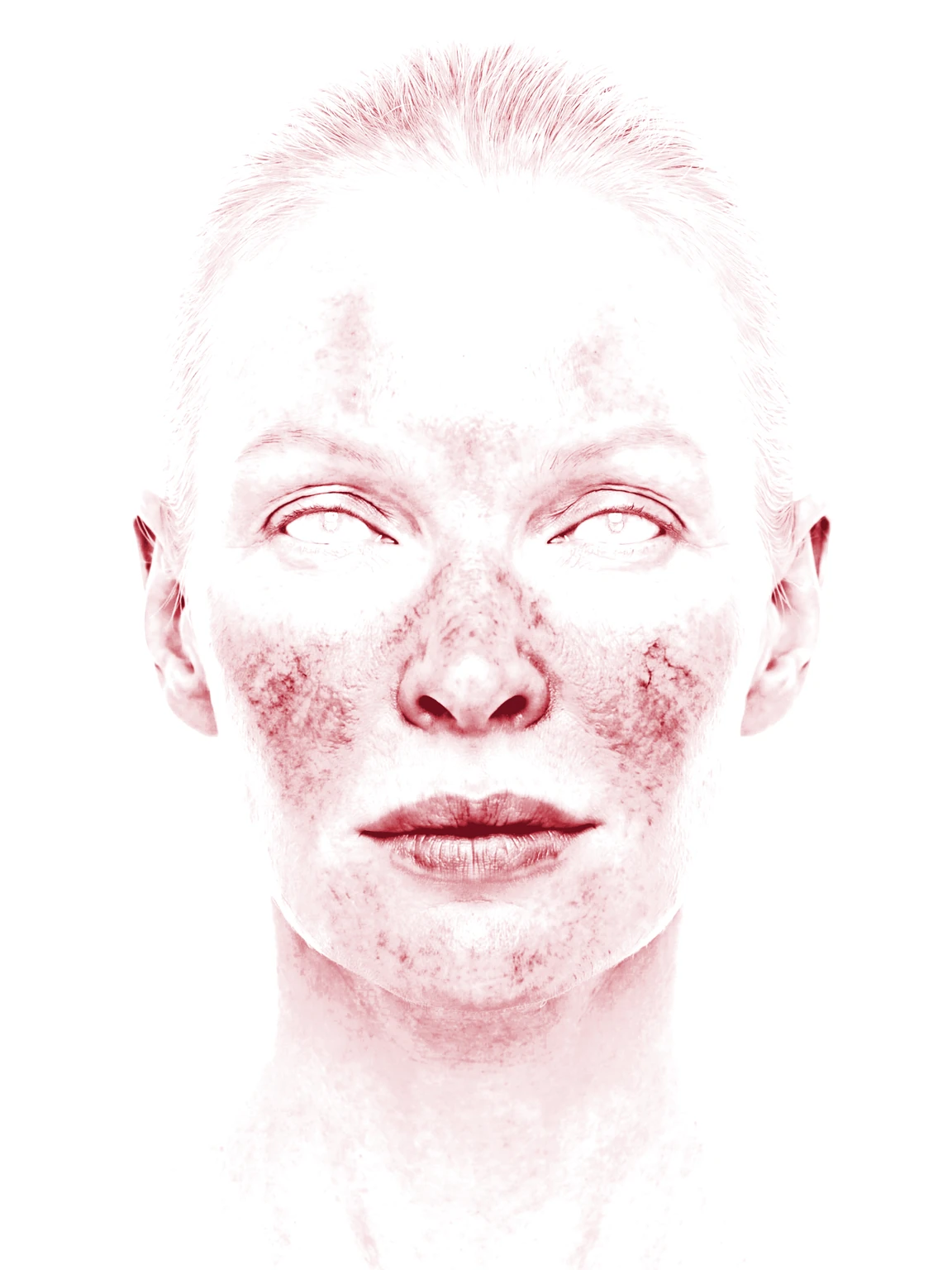
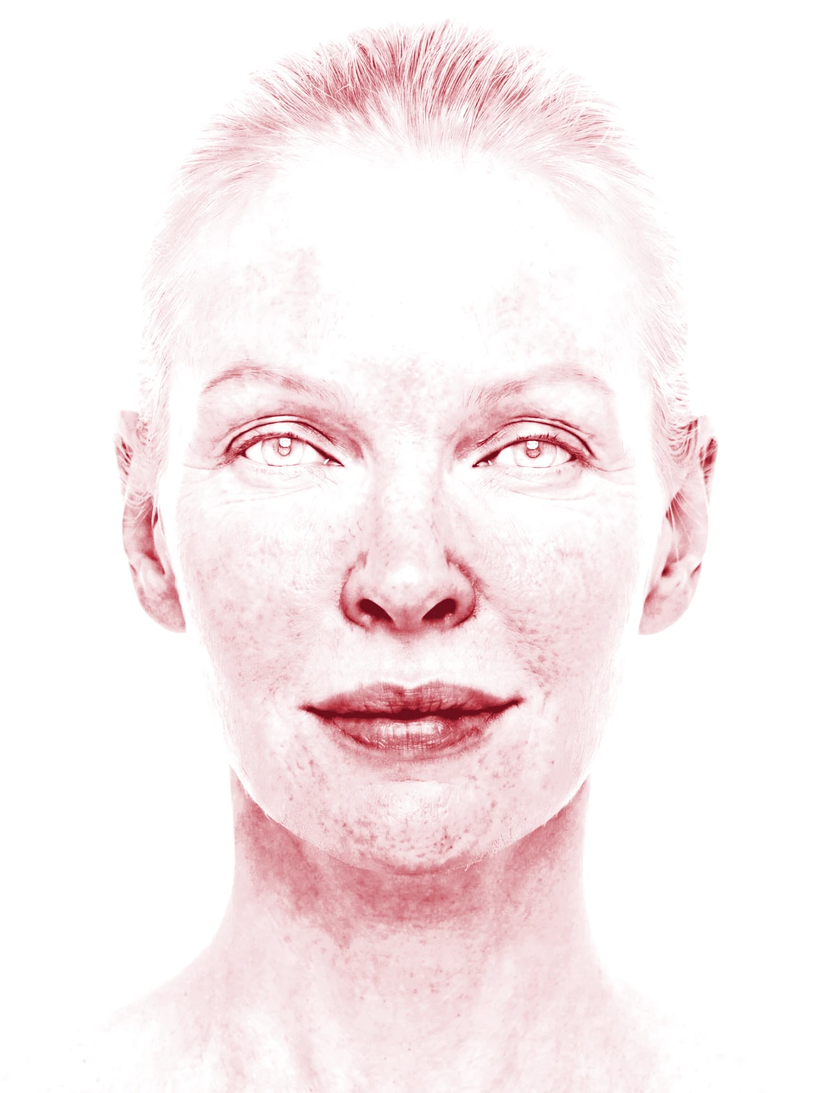
Illustrative demo with synthetic data. Learn more
Frequently asked questions
Filters change only how the photograph is displayed; the stored photograph keeps its original pixels. Annotations and lesion markers stay unfiltered on top, and each clinician and device chooses its own filter.
Every image in a view shares one filter and one strength, so a difference between visits is shown as it is. Lighting and white balance changes show as clearly as a change in the skin, so matched lighting compares best.
No. Original and annotated image downloads and PDF reports use the unfiltered photographs, whatever filter is on screen; only Custom Export and Quick Export render it, labeled with the filter name.
No. Cross-polarized, UV, and dermoscopic imaging depend on how the skin is lit at capture and remain separate modalities. A filter works with the color and tone a standard photograph already recorded.
Dermi Atlas is a clinical documentation platform; it does not make diagnoses. The displayed image is the only output of a filter, and every clinical assessment of a filtered photograph is made and recorded by the clinician.
Try image filters in Dermi Atlas
The demo runs in a browser; the free trial runs on the computers your practice already owns.
Resources and further reading
Related pages
Plans, pricing, and free trial
Atlas Cloud Demo
- No time limit
- No credit card
- Nearly every feature, with photos you upload
- Runs in a browser, nothing to install
- A free Dermi Cloud Services account to sign in
- Not for real patient data
- Data cleared at least weekly
Dermi Atlas Professional
- Base plan per deployment
- First 30 days free
- Additional clinician seat$59 USD / month
- Additional assistant seat$12 USD / month
- Every feature, with real patient records
- Runs on computers the practice already owns
- Technical support at no extra charge
Synthetic Data Notice
All demonstrations, screenshots, and media on this page use synthetic data only. No real patient information is shown.
The following are synthetic and do not correspond to real patients:
- All human faces and individuals are synthetic and do not represent real people
- All clinical and medical images, including photographs and scans, are synthetic or simulated
- All patient names, dates, identifiers, and other details are fictional
- All clinical notes and documentation are sample content for demonstration only
Media is provided solely to illustrate platform functionality and workflows.